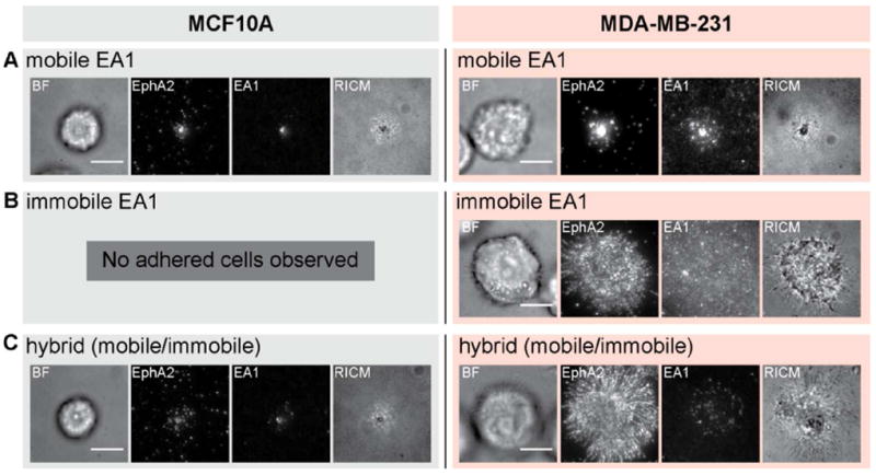
Figure 3.

Comparison between MCF10A and MDA-MB-231 cells placed on substrates with three different ephrin-A1 configurations. A) Normal EphA2 transport is observed for both MCF10A and MDA-MB-231 cells on mobile ephrin-A1 surfaces. B) On entirely immobile ephrin-A1 surfaces, and after rinsing steps during fixation and permeabilization processes, only MDA-MB-231 cells adhere. C) MCF10A cells adhere to hybrid surfaces, containing both mobile and immobile ephrin-A1, and EphA2 transport is normal. In contrast, MDA-MB-231 cells exhibit jammed phenotypes on these surfaces. Scale bars are 10 microns.
