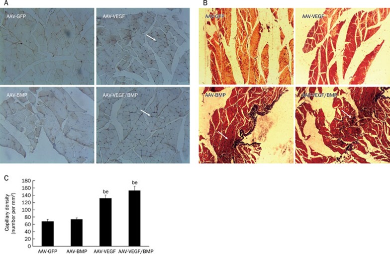
Figure 8.
Histological assessment. (A) Representative images of rabbit muscle tissues using immunostaining for CD34 (capillaries were indicated as arrows). (B) Representative images of rabbit muscle tissues using von Kossa staining for calcium deposits. The calcium deposits were stained as black (indicated as arrows). (C) Capillary density in rabbit muscle tissues. The number of capillaries was counted in twenty different fields of one muscle section, and capillary density was calculated. Data are shown as mean values±SEM from three independent experiments. (magnification×200). bP<0.05 vs AAV-GFP group. eP<0.05 vs AAV-BMP group.

