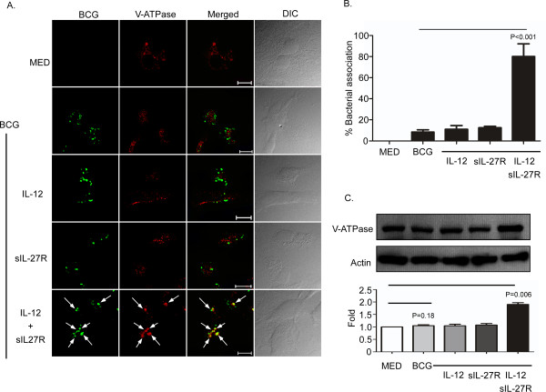
Figure 4.
Treatment of BCG-infected macrophages with IL-12 and sIL-27R increases expression of V-ATPase. Macrophages were treated with IL-12, sIL-27R, or their combination for 6 h and then infected with SYTO-9®-stained BCG for 48 h. (A) The cultures were subsequently fixed with 4% PFA, permeabilized, and stained with anti-VATPase H antibody (red). Representative images from three experiments are shown. Arrows indicate the association of V-ATPase with BCG. (B) The percent bacterial association was calculated as described in the Methods section. These data are representative results from three independent experiments. (C) Macrophage cultures were lysed to collect whole-cell lysates for immunoblot analysis (upper panel). V-ATPase or actin was labeled as described in the Methods section. An image representative of two experiments is shown. The ratio of V-ATPse/actin band intensity was expressed relative to medium alone (lower panel). Graphical data plotted are the result of two combined experiments. (B, C) A student’s t test was used to establish statistical significance in the 95% confidence interval between individual sample groups as indicated.

