Fig. 5.
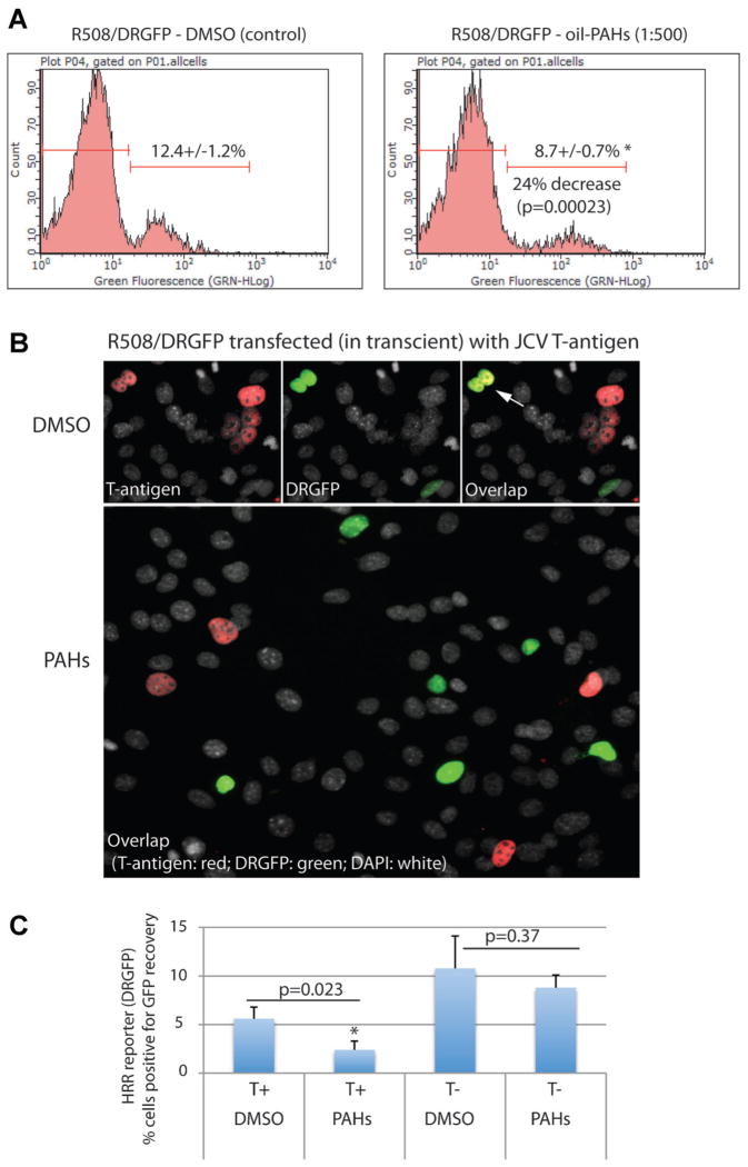
Effects of oil-PAHs and JCV T-antigen on HRR. Part A: R508 cells expressing HRR reporter plasmid (R508/DRGFP) were transfected in transient with DNA damaging pCβA-Sce expression vector and the cells were cultured in the presence of 10%FBS ± oil-PAHs (1:500) for 96 h. The cells capable of reconstituting GFP function were analyzed by flowcytomentry. The results were collected from three separate experiments, in duplicate (n = 6). Part B: Cytofluorescent detection of HRR in R508/DRGFP cells expressing JCV T-antigen. The cells were transfected (nucleoporation; Amaxa) with pCDNA3/JCV-T/Zeo (Trojanek et al., 2006a) and with pCβA-Sce (Pierce et al., 1999) expression vector. Following 24 h of recovery after transfection, the cells were treated either with oil-PAHs (1:500) or with DMSO (control). JCV T-antigen was detected using anti-SV40 T-antigen mouse monoclonal antibody (Calbiochem; red fluorescence). DRGFP fluorescence is indicated green, and total nuclei are labeled with DAPI (white fluorescence). Arrow indicates two nuclei in which T-antigen expressing cells are positive for HRR-mediated reconstitution of DR-GFP otherwise the majority of T-antigen expressing cells do not express green. Part C: Quantification of the results depicted in part B. The results were collected from three separate experiments, in duplicate in which 1,000 cells per experiment were counted in at least 50 randomly selected microscopic fields. P-values are indicated above the compared samples (paired Student’s t-test P ≤ 0.05).

