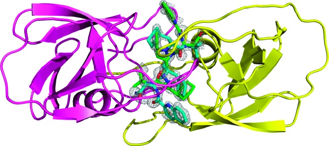
Figure 1.

Dimeric structure of HIV-1 protease (ribbon/loop rendering) viewed parallel to the 2-fold symmetry axis. The electron density 2Fo−Fc map (contour level at 1.0σ) of the catalytic channel of HIV-1 protease shows the double orientation (green and cyan sticks) of SQV.
