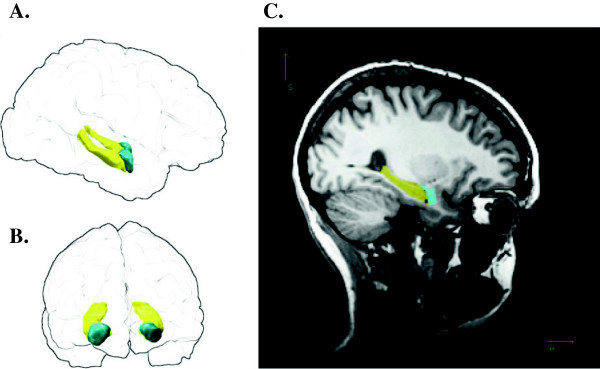
Figure 1.

Parcellation of hippocampus and amygdala in a representative participant (female, right hippocampal volume ranked 15/30). Left: “glass brain” renderings showing three-dimensional volumes of right and left hippocampi (yellow) and right and left amygdalae (cyan) viewed from participant’s right (A) and front (B); the outline of the pial surface is shown in black. C. Right labelled voxels overlaid on a T1 image, sagittal section passing through right hippocampus (yellow) and amygdala (cyan).
