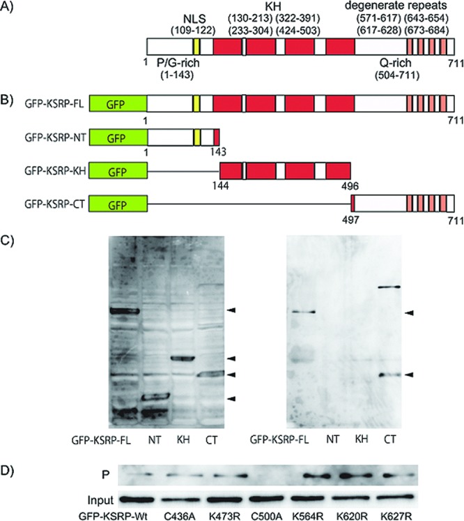
Figure 4.

Structure of KSRP/FUBP2 and determination of the GO-Y086-binding site. (A) The domain structure of KSRP/FUBP2. NLS: nuclear localization signal. KH: KH RNA-binding domain. Degenerate repeats: DYTKAWEEYYKK degenerate repeats. P/G-rich: proline/glycine-rich domain. Q-rich: glutamine-rich domain. (B) Structure of the GFP-tagged deletion mutants of KSRP/FUBP2. (C) Western blot analysis of the GO-Y086-bound mutant using anti-GFP antibody. HEK-293T cells were transfected with different GFP-tagged deletion mutants, as shown in panel B. The mutants in the cell lysates were tested for their ability to bind to GO-Y086. Left panel: total cell lysate. Right panel: precipitate. (D) Western blot analysis of GO-Y086-bound point mutants of KSRP/FUBP2 using anti-GFP antibody. Input: total cell lysate. P: precipitate. GFP-KSRP-Wt: GFP-tagged KSRP wild-type.
