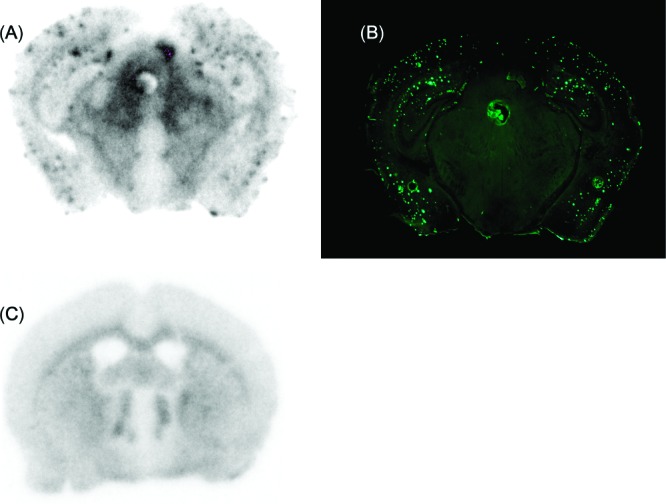
Figure 3.

Labeling of β-amyloid plaques in vivo was visualized by autoradiography ex vivo with [18F]17 in sections of Tg2576 mouse brain (A). The same section was also stained with thioflavin-S (B). Wild-type mouse brain showed no β-amyloid plaques (C).
