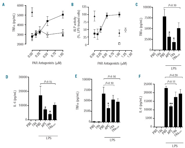
Figure 2.
FXa inhibits LPS-induced pro-inflammatory cytokine production from myeloid cells via PAR2 activation. (A) PBMC were exposed to PAR1 (FR131117;○) or PAR2 (GB83;■) antagonists for 30 min prior to FXa (20nM) co-incubation for 3 h, followed by LPS treatment (50 ng/mL; 18 h). TNFα was then measured by ELISA. LPS-induced TNFα in the absence of FXa (▲) and LPS-induced TNFα in the presence of APC/GB83 is also shown (♦). (B) THP-1 cells were exposed to GB83 (0.125–1 μM) for 30 min prior to incubation with FXa (20nM;■) or APC (20nM;♦) for 3 h. Similarly, THP-1 cells were exposed to FR131117 (1.25 μM) for 30 min prior to incubation with FXa (20nM;○) for 3 h. Cells were treated with LPS (500 ng/mL; 4 h) and TNFα secretion was detected using HEK Blue TNFα reporter cells. LPS-induced TNFα in the absence of FXa or PAR antagonists is shown (▲). GB83 did not induce TNFα production in the absence of LPS (□). Murine bone marrow-derived macrophages were isolated from wild-type (C and D) and PAR2−/− (E and F) BALB/c mice and exposed to APC/FXa/FXaDEGR (all 20 nM) for 3 h prior to addition of PBS or LPS (20 ng/mL) for 18 h. Murine TNFα (C and E) and IL-6 (D and F) were determined by ELISA and the mean ± SD from three independent experiments is shown.

