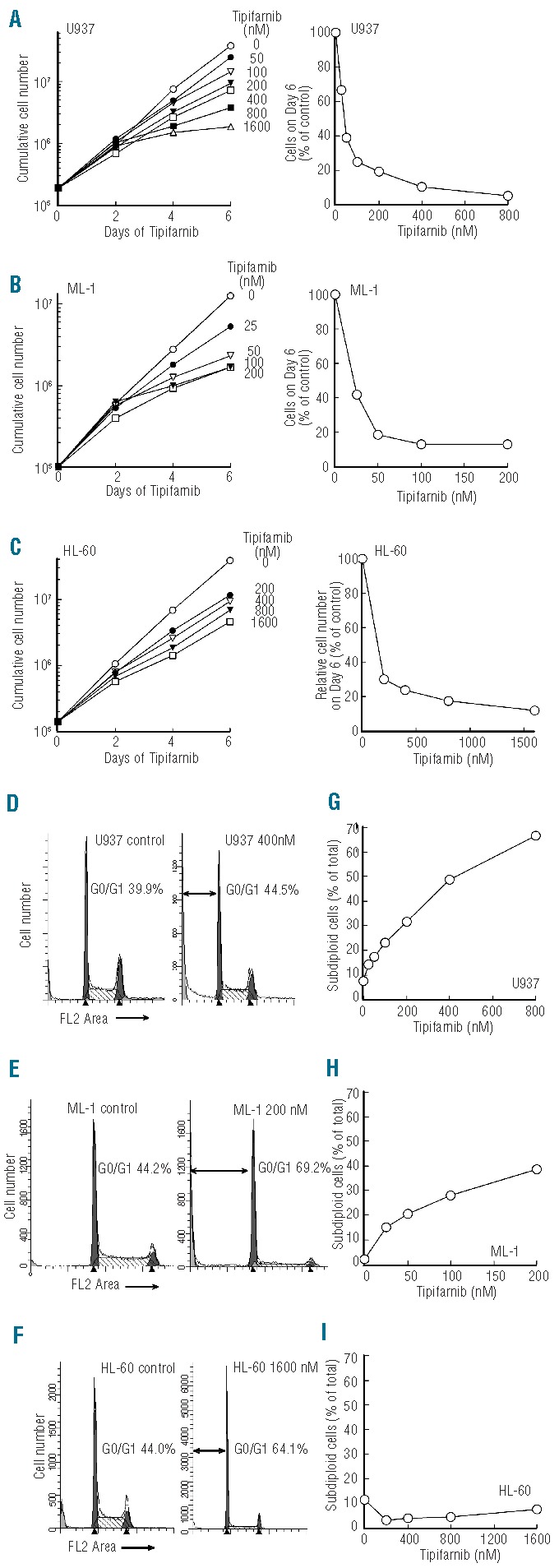
Figure 1.

Tipifarnib inhibits proliferation and induces apoptosis in AML cell lines. (A–C) Aliquots containing 1−2×10⁵ HL-60, U937, and ML-1 cells/mL were treated with the indicated concentrations of tipifarnib or diluent continuously, with cells being counted and diluted into fresh medium with drug every 2 days. Graphs on the left indicate cumulative cell number/mL after taking into account dilution. Graphs on the right show cell number on Day 6 relative to diluent-treated controls. (D–F) DNA histograms of cells treated with diluent or the indicated tipifarnib concentration for 6 days. Double-headed arrows indicate events with <2n DNA content. G-I, percentage of subdiploid events observed on Day 6 after analysis of FL2 height as illustrated in Online Supplementary Figure S2A. Results in all panels are representative of at least 3 independent experiments.
