Figure 5.

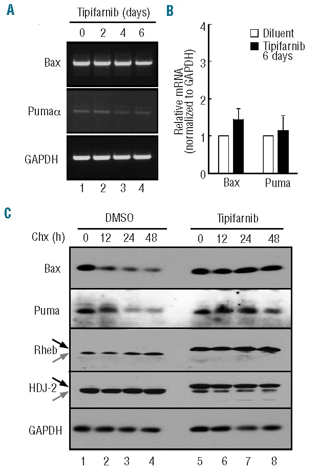
Posttranslational stabilization of Bax and Puma. (A and B) after U937 cells were treated with 800 nM tipifarnib for the indicated time in the presence of 5 μM Q-VD-OPh, a broad spectrum caspase inhibitor, RNA was isolated and subjected to semiquantitative (A) or quantitative RT-PCR (B) using GAPDH as a loading control. (C) After treatment for 3 days with diluent (lanes 1–4) or 800 nM tipifarnib (lanes 5–8), cells were supplemented with 30 μM cycloheximide for 0–48 h in the continued presence of diluent or tipifarnib, then subjected to SDS-PAGE followed by immunoblotting with antibodies that recognize the indicated antigens. GAPDH served as a loading control. The similar levels of Bax and Puma in lanes 1 and 5 are consistent with time course experiments in which a 6-day tipifarnib treatment up-regulated Bax or Puma (Figure 4A) but a 3-day treatment (Figure 4C) did not.
