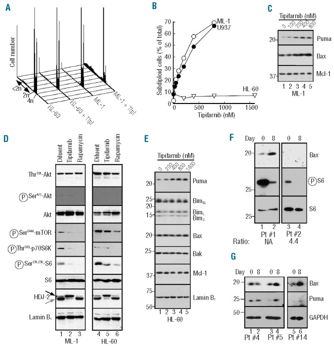
Figure 7.

Correlation between tipifarnib-induced Bax upregulation and apoptosis. A. After ML-1 cells were treated for 6 days with the indicated tipifarnib concentration, samples were stained with propidium iodide and subjected to flow microfluorimetry. B. Summary of subdiploid events observed after U937, ML-1 or HL-60 cells were analyzed as illustrated in panel A. C. After ML-1 cells were treated with diluent (lane 1) or tipifarnib at 100, 200, 400 or 800 nM (lanes 2–5, respectively) for 6 days, whole cell lysates were subjected to SDS-PAGE followed by immunoblotting with antibodies to the indicated antigen. D. After ML-1 (lanes 1–3) or HL-60 (lanes 4–6) were treated for 48 h with 0.1% DMSO (lanes 1, 4), 800 or 1600 nM tipifarnib (lanes 2 and 5, respectively) or 100 nM rapamycin (lanes 3, 6), whole cell lysates were subjected to SDS-PAGE followed by immunoblotting with antibodies to the indicated antigen. For each antigen, all lanes were derived from corresponding signals on a single piece of x-ray film but were rearranged for clarity. E. After HL-60 cells were treated for 6 days with diluent (lane 1) or tipifarnib at 200, 400, 800 or 1600 nM (lanes 2–5, respectively), whole cell lysates were subjected to SDS-PAGE followed by immunoblotting with antibodies to the indicated antigens. The nucleoskeletal protein lamin B1 served as a loading controls in panels (D and E). (F and G). Aliquots containing 5 × 10⁵ marrow mononuclear cells harvested prior to treatment (Day 0) or after 14 doses of tipifarnib (on Day 8 before further drug administration) on a phase II trial of tipifarnib (Days 1–14) + etoposide (Days 1–3 and 8–10)28 (F) or a phase II trial of single-agent tipifarnib27 (G) were subjected to SDS-PAGE followed by immunoblotting for the indicated antigens. Number below panel in F indicates RasGRP1:aprataxin ratio previously reported for the corresponding mRNA sample.28 NA: not assayed due to lack of RNA sample.
