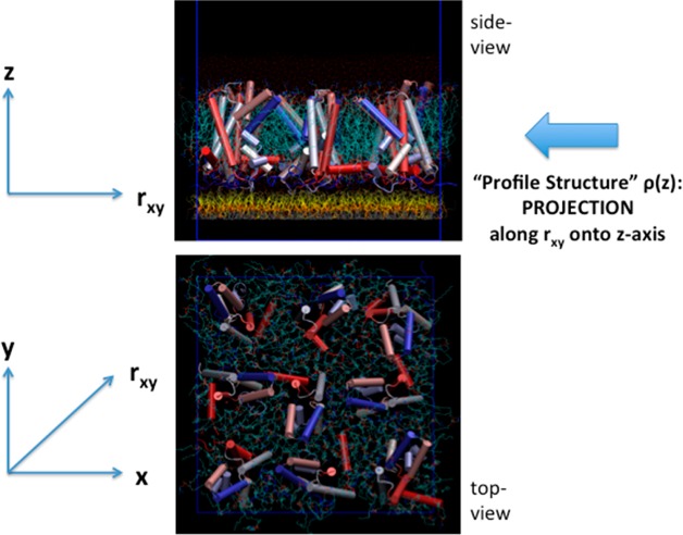
Figure 1.
Molecular graphics representation of a small 3 × 3 ensemble of the vectorially oriented VSD molecules (α-helices represented as solid cylinders) solvated by a POPC bilayer (turquoise) tethered to the surface of a solid inorganic substrate (not shown) below the monolayer of tethering chain molecules (yellow) in the side view. The “profile structure” arises from the projection of the 3-D structure of the membrane along rxy vectors parallel to the plane onto the z-axis normal to the plane of the membrane. The VSD molecules have been rotated randomly with respect to one another about the z-axis in the top view. However, unlike in the top view shown, experimentally, the VSD’s exhibit only glass-like order in the x–y plane, even at this low average area/VSD of ∼1400 Å2 approximating that of the experimental situation.

