
Figure 2.
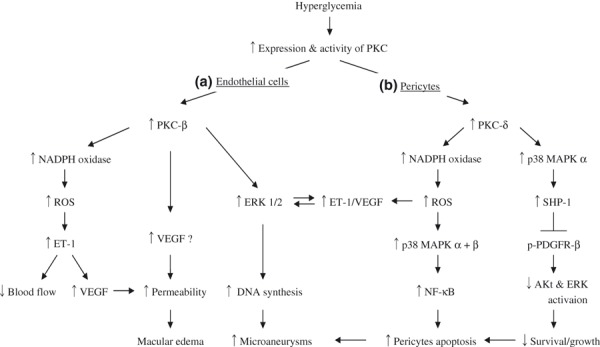
Outline for mechanisms of diabetic retinopathy. (A) Activation of protein kinase C (PKC)‐β in retinal endothelial cells contributes to increased vascular permeability and formation of microaneurisms. (B) Activation of PKC‐δ induce retinal pericytes apoptosis through activation of nuclear factor‐κB (NF‐κB) by oxidative stress and activating Src homology‐2 domain containing phosphatase‐1 (SHP‐1) to inhibit platelet‐derived growth factor’s (PDGF) survival actions. Akt, protein kinase B; ERK, extracellular signal regulated kinase; ET‐1, endothelin‐1; MAPK, mitogen activated protein kinase; NADPH, nicotinamide adenine dinucleotide phosphate; PDGF, platelet‐derived growth factor; ROS, reactive oxygen species; TGF‐β, transforming growth factor‐β; VEGF, vascular endothelial growth factor.