Figure 1.

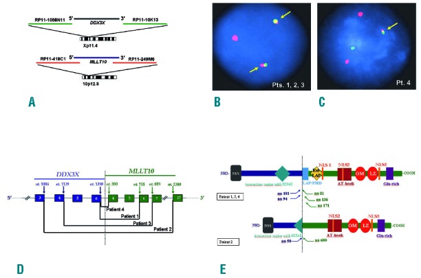
Cytogenetic and molecular characterization of DDX3X-MLLT10 fusions. (A) Double color double fusion FISH assay for DDX3X and MLLT10. (B) FISH showed 2 fused signals in Patients 1, 2 and 3 indicating a balanced translocation (arrows). (C) FISH showed one fused signal in patient 4 indicating an unbalanced translocation (arrow). (D) Schematic representation of DDX3X and MLLT10 breakpoints in the 4 DDX3X-MLLT10 positive cases (arrows). Nucleotide numbers refer to GenBank accession: NM_001356.3 for DDX3X and NM_004641.3 for MLLT10. (E) Putative fusion protein structure. At N terminal DDX3X retained a NES domain in all. Three patients retained the entire EIF4E interacting motif and 1 only half. At C terminal at least 1 NLS, the AT-hook and the OM-LZ domain were retained in all. Pt: patient; Pts: patients; nt.: nucleotide; aa: amino acid; NES: Nuclear Exporting Signal; NLS: Nuclear Localization Signal; LAP/PHD: Leukemia Associated Protein / Plant Homeo Domain; Ext-LAP: Extended LAP; OM-LZ: Octapeptide Motif-Leucine Zipper; Gln: Glutamine.
