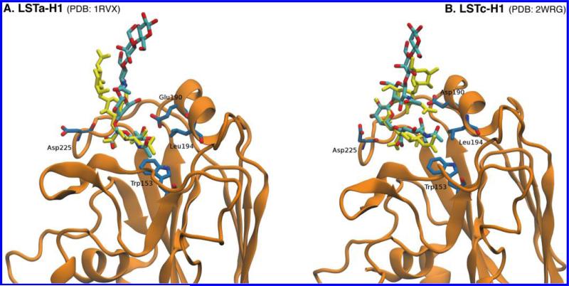
Figure 6.
Comparison of the MD -generated LSTa (A) and LSTc (B) conformers (structures with the smallest rmsdexp value, rmsdexp min) and crystal structures of LSTa and LSTc bound to hemagglutinin from H1. The conformers generated via MD simulations are colored cyan (C atoms) and red (O atoms), while the conformers determined by X-ray crystallography are colored yellow. The rmsd values between the MD simulation-generated structures and the cocrystallized glycans are 6.2 and 6.5 Å for LSTa and LSTc, respectively.

