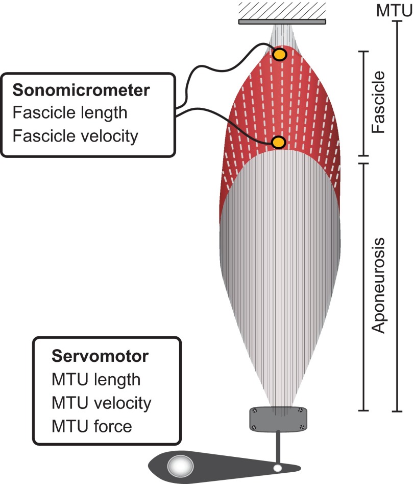
Fig. 1.
Schematic diagram of the in vitro preparation. The regions corresponding to the fascicle, aponeurosis and MTU are indicated. The muscle is stimulated maximally through its nerve. Sonomicrometry is used to measure fascicle length and velocity. A dual-mode servomotor is used to measure MTU force, length and velocity. The ratio of MTU velocity to fascicle velocity is used to characterize the muscle's AGR.

