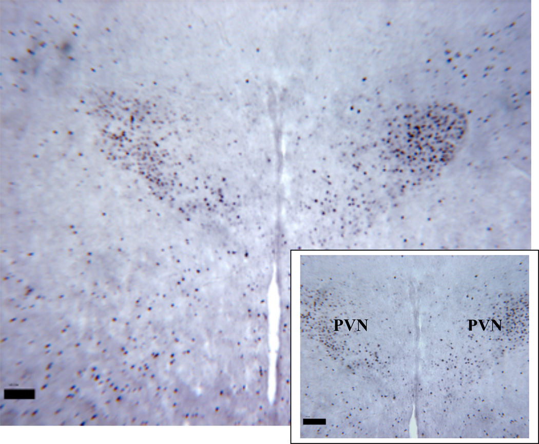
Figure 1.
Low and high magnification of a typical coronal section of PVN from a Fos positive (black dots represent counts of Fos production) control animal. Water was withheld for 24 hours prior to tissue collection to induce Fos expression. One such control animal was performed with every group to verify DAB staining intensity (PVN=paraventricular nucleus).
Magnification bar = 100 µm

