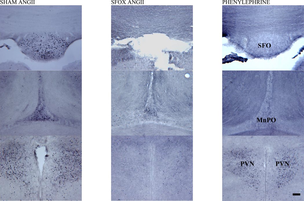
Figure 2.
Representative coronal sections from subfornical organ (SFO; top row), dorsal median preoptic nucleus (MnPO; middle row) and paraventricular nucleus (PVN; bottom row) in animals following 2 hours of intravenous infusion of AngII in sham (first column) and SFOx (second column) animals, as well as phenylephrine (final column) treated control rats. Midline structures of the lamina terminalis are labeled in the third column as SFO and MnPO, as well as the bilaterally paired areas of the hypothalamic PVN.
Magnification bar = 100 µm.

