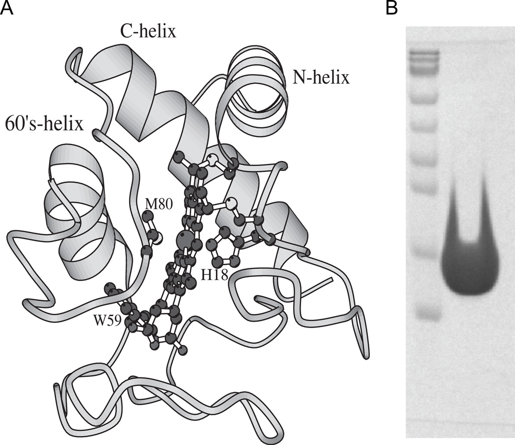
Fig. 1.
(A) Structure of Cyt c (PDB ID: 1HRC). The protein consists of three α-helices and three Ω-loops. The figure also shows the covalently linked heme, its two axial ligands His18 and Met80, and the single Trp residue at position 59 which fluoresces in the unfolded state. The structure was generated using the MOLSCRIPT59 program. (B) SDS/PAGE of Cyt c used in this study. Lane 1 shows the protein molecular weight markers (bottom to top: 10, 17, 26, 34, 43, 56, 72, 95, and 130 kDa respectively). Lane 2 was overloaded with Cyt c, whose expected molecular weight is 12.4 kDa, to see whether or not other protein impurities are present in the sample.

