
Figure 1.
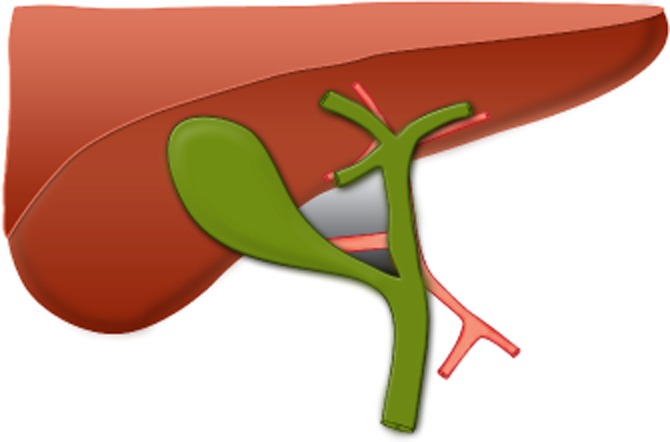
Landmarks of the hepatobiliary triangle. The grey area shows the hepatobiliary triangle, bounded by the common hepatic duct, cystic duct and inferior border of the liver. The smaller dark grey area shows Calot's triangle, bounded by the cystic duct and cystic artery and common hepatic duct