
Figure 2.
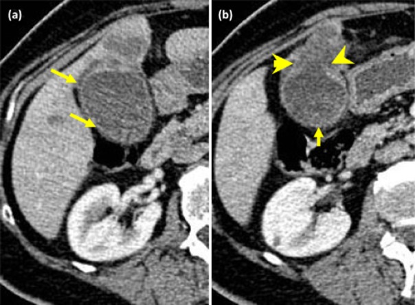
Computed tomography (CT) scans from patients with biopsy-proven gall bladder cancer. (a) Circumferential enhancement of the mucosa with focal wall thickening (small arrows). (b) Circumferential enhancement of the mucosa with focal thickening (small arrow) and breach of the mucosa (arrowheads) with invasion into segment 4b of the liver (contrast with the figure showing XGC)