
Figure 1.
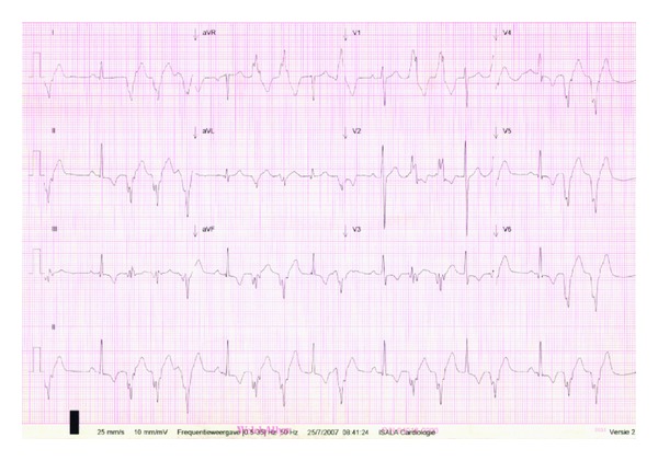
Twelve lead ECG showing ventricular bigeminy, doublet PVC's with an RBBB configuration with an RS interval of >100 msec in the precordial leads, and a pseudodelta wave configuration suggesting an epicardial origin of the ventricular ectopy.