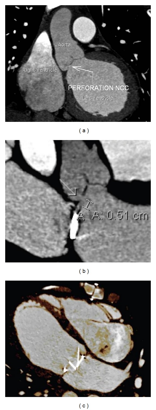
Figure 3.

Cardiac gated computed tomography. (a) Large and (b) magnified oblique reformatted images of the left ventricular outflow tract and aortic valve demonstrate a clearly visible 5 mm defect in the non-coronary cusp of the aortic valve. (c) Four-chamber orientation shows the asymmetric movement of the bileaflet mechanical mitral valve due to the regurgitant jet originating from the aortic valve defect. Supplementary CINE file online shows 3D rendering of paradoxical mitral valve motion (see supplementary CINE file).
