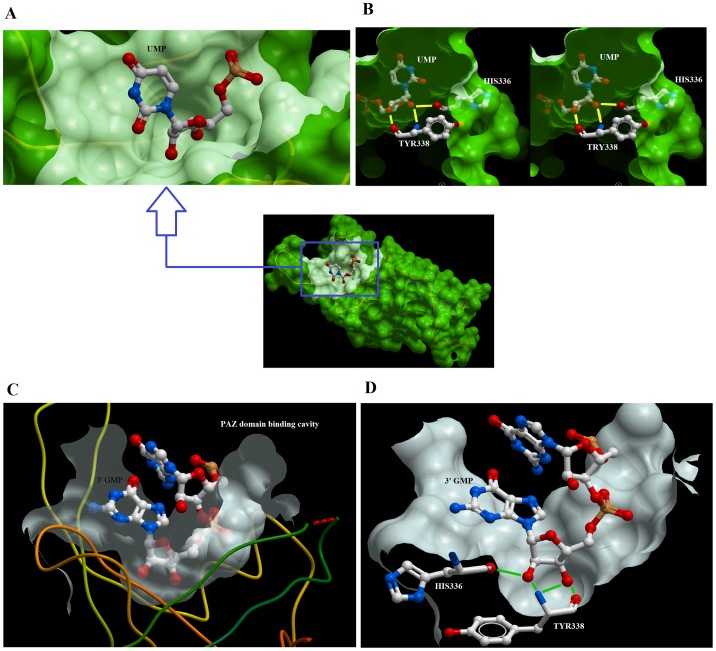
Figure 5. Surface representation of the interaction of nucleotides with PAZ domain of Human Ago2.
Accessible surface representation of the human EIF2C2/Ago2 PAZ domain (A) showing the binding cavity of the PAZ domain bound with UMP at its 3′-terminal. Stereoview showing the hydrogen bonding of the ribose sugar with the side chains of HIS110 and TYR112 (B). The binding cavity of the EIF2C2/Ago2 PAZ domain bound with GMP at its 3′-terminal (C). Hydrogen bonds between the ribose of GMP and the side chains of HIS110 and TYR112 are shown in green (D). The coordinates of interaction were derived from PDB ID 4EI1 and 4F3T. The figure was generated using Molsoft.

