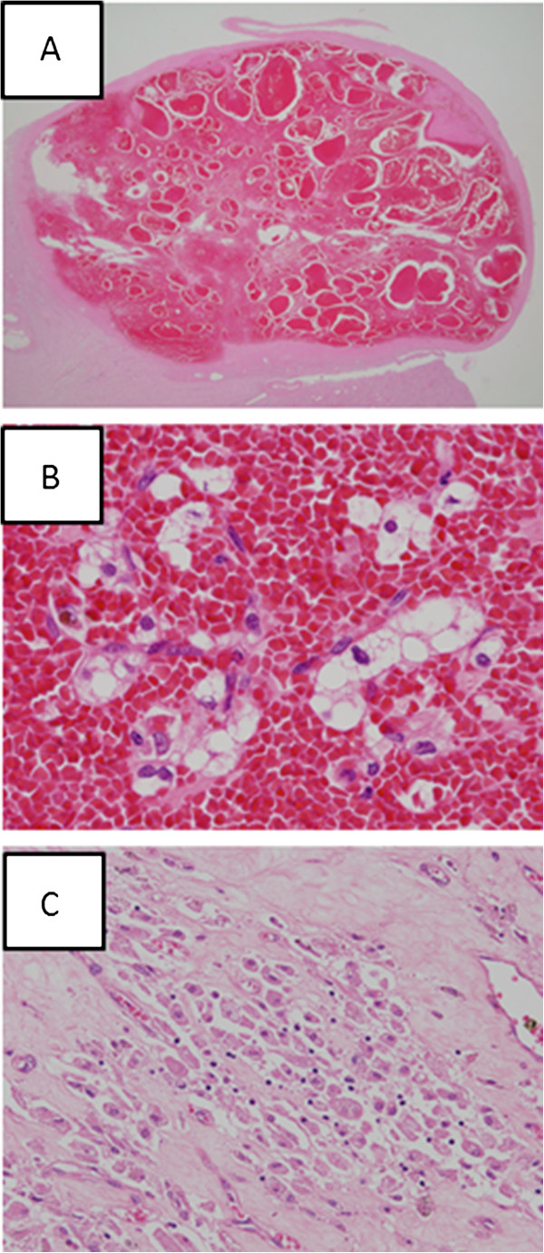
Figure 3.
Histological findings of clear cell renal cell carcinoma treated with stereotactic radiotherapy. (A) The tumor at low magnification shows markedly hemorrhagic findings within a fibrous capsule. (B) A high-power view demonstrating clustered, degenerative cancer cells with abundant, clear and/or vacuolated cytoplasm and round-to-ovoid or irregularly-shaped nuclei with a hemorrhagic background. (C) Aggregation of foamy macrophages in the fibrotic stroma.

