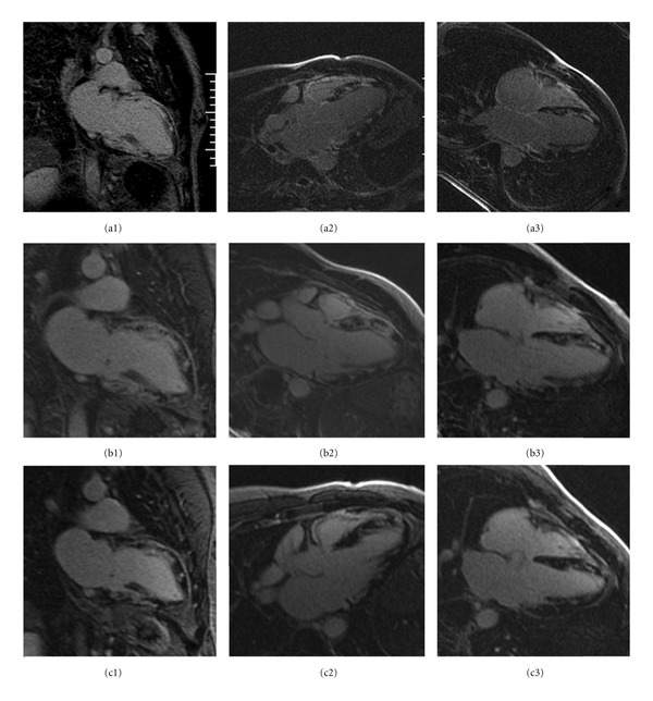
Figure 2.

Delayed gadolinium hyperenhancement images. DHE-CMR pattern of patchy delayed hyperenhancement secondary to giant cell myocarditis or a severe form of cardiac sarcoidosis. Patchy hyperenhancement in left ventricular wall and diffuse enhancement in right ventricular wall in the initial exam (a1, a2, and a3) has improved to coalescence of hyperenhancement in seven weeks on cyclosporine and prednisone (b1, b2, and b3). The nine-month exam (c1, c2, and c3) shows a stabilization of change in hyperenhancement as compared to the seven-week exam.
