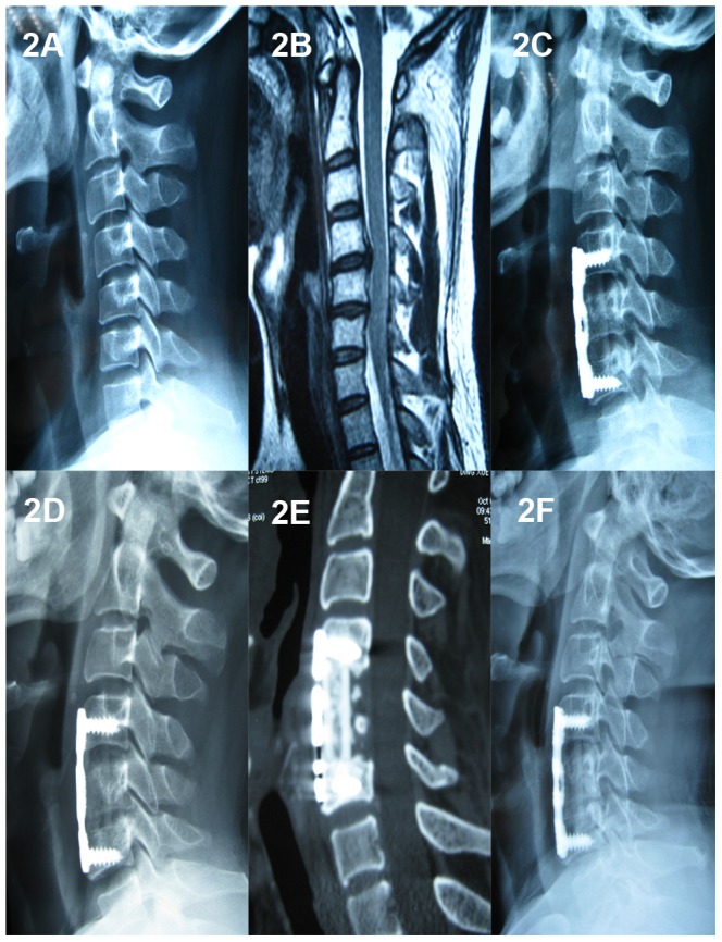
Figure 2. A 36-year-old male who underwent 1-level corpectomy with a nano-hydroxyapatite/polyamide66 cage used for cervical reconstruction.

The preoperative cervical X-ray film (2A) and MRI scan (2B) show the spinal cord compression resulting from C4/5 and C5/6 disc herniations. The immediately postoperative lateral X-ray (2C) shows C5 corpectomy and the n-HA/PA66 cage used for reconstruction, and an obvious radiolucent gap can be observed between the cage and the endplates. The lateral X-ray film (2D) shows no obvious radiolucent gap, and the 3D-CT (2E) scan shows the autogenous bone granules filling the cage and achieving bony fusion with adjacent endplates at the 1-year follow-up. A lateral X-ray film (2F) at the final follow-up (four years and eight months) shows satisfying bony fusion and no obvious migration or subsidence.
