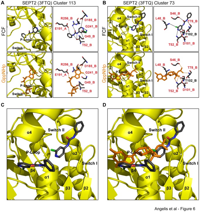
Figure 6. In silico binding of FCF to two distinct sites of the nucleotide-binding pocket of SEPT2-GppNHp.
(A) Ribbon and stick diagrams show the orientation and atomic interactions of a representative pose of FCF bound to the GppNHp-bound crystal structure of SEPT2 (PDB: 3FTQ) from the cluster of conformations (113 out of 250) with the lowest binding free energy. Red text denotes amino acids and their corresponding protomers that interact with both FCF and GDP. (B) Ribbon and stick representations depict the position and atomic bonds of a representative pose of FCF bound to the GppNHp-bound crystal structure of SEPT2 (PDB: 3FTQ) from the cluster of conformations (73 out of 250) with the second lowest binding free energy. Red letters denote amino acids that interact with both FCF and GDP. (C and D) Ribbon representations show the two conformations of FCF superimposed with the structure of SEPT2 in the absence (C) and presence of GppNHp (D). Note the lack of overlap between the two FCF conformations, which overlap with distinct portions of the GppNHp molecules (guanosine base vs. gamma phosphate).

