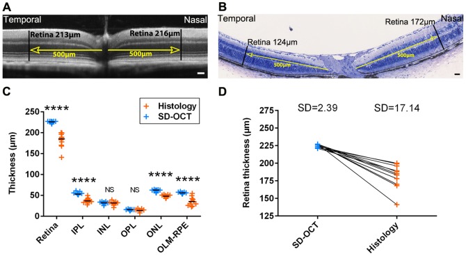
Figure 1. Retinal layer thickness measures in C57BL/6JRj wild-type mice by SD-OCT and histology.
Retinal thickness in nasal and temporal sides in SD-OCT image (A) and in corresponding histological section (B). (C) Measures of retinal layers thickness by SD-OCT and histology in C57BL/6JRj mice, n = 11, Mann Whitney test. (D) Retinal thickness evaluated by SD-OCT and histology in C57BL/6JRj mice. Each pair of point represents the whole retina thickness of the same eye measured with SD-OCT (blue dots) and histology (orange dots). IPL: inner plexiform layer, INL: inner nuclear layer, OPL: outer plexiform layer, ONL: outer nuclear layer, OLM: outer limiting membrane, RPE: retinal pigmented epithelium. SD: Standard Deviation. Scale bars: 50 µm.

