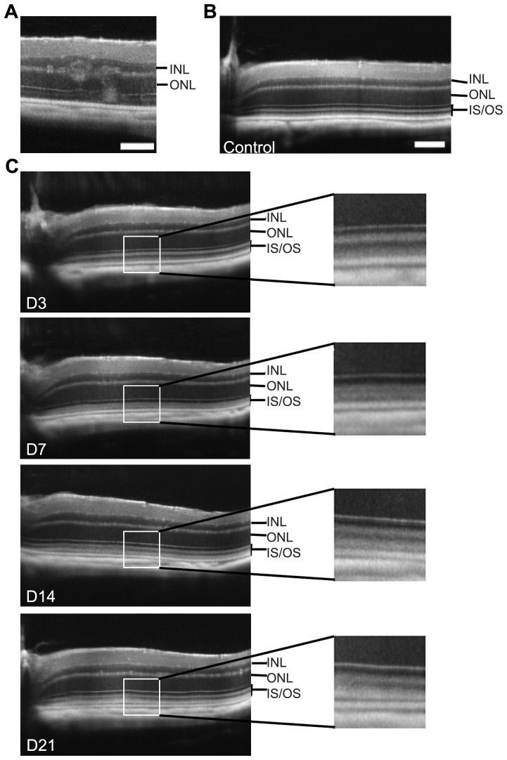
Figure 4. SD-OCT imaging in other pathological models: rd8 mutation and light-challenge.
(A) Typical ocular lesions of rd8 mutation in crb1 gene (C57BL/6NRj mice in which presence of the rd8 mutation was confirmed by genotyping). (B–C) SD-OCT follow-up of the outer retina during a light-challenge in C57BL/6JRj mice. Control unexposed three month-old mouse has a normal appearance with 4 bands of different reflectance corresponding to the PR segments (B). Mice were then exposed to light during 4 days as described in the “methods” section and the retina was imaged by SD-OCT at day 3 (D3), 7, 14 and 21 after starting the illumination (C). The light-challenge leads to a temporary abolition of the distinction between the two bands forming the outer segment, with a peak at D7 (right panels: enlargement of the area enclosed by a white box on the left view). INL: Inner Nuclear Layer, ONL: Outer Nuclear Layer, IS: Inner Segments, OS: Outer Segments. Scale bars: 50 µm.

