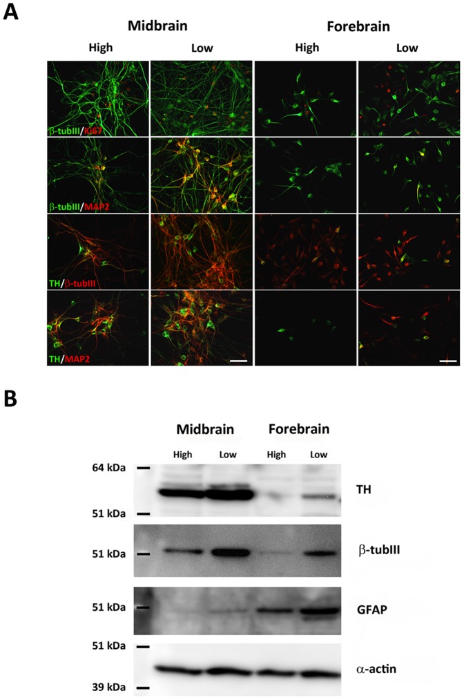
Figure 4. Characterization of differentiated cells.
Double immunofluorescence staining was performed for histological characterization of differentiated cultures (A). A small number of cells in both midbrain and forebrain-derived cultures co-expressed the marker of proliferating cells, Ki67, and the early neuronal marker β-tubulin III (β-tub III). The density of β-tub III-ir cells co-expressing the mature neuronal marker microtubule associated protein 2ab (MAP2) was very high for midbrain cultures, particularly when differentiated at low oxygen tension. In forebrain-derived cultures, only few β-tub III-ir/MAP2-ir cells were detected at low oxygen and even fewer at high oxygen. The density of cells co-expressing tyrosine hydroxylase (TH) and β-tub III or MAP2 was highest for midbrain cultures grown at low oxygen, and lowest for forebrain cultures at high oxygen. Scale bar = 50 µm. Western blotting for TH, β-tub III, and the astroglial marker glial fibrillary acidic protein (GFAP) revealed marked differences depending on both cell origin and oxygen tension (B). The expression of all these markers was higher in cultures differentiated at low oxygen. The level of β-tub III and TH protein was highest for midbrain-derived cells, whereas the highest level of GFAP was detected for forebrain-derived cells.

