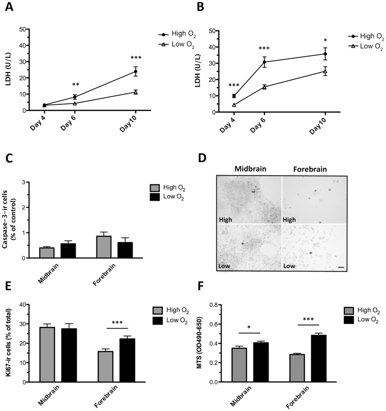
Figure 5. Effects of oxygen on cell death and cell proliferation.
Assessment of cell death, cell proliferation and cell viability in midbrain and forebrain NSC cultures differentiated (sequential addition of FGF8, Shh, GDNF, and Forskolin) for 10 days at high (20% O2) or low oxygen tension (3% O2). Level of lactate dehydrogenase (LDH) in conditioned culture medium from differentiating midbrain (A) and forebrain (B) cells. For both cell types, significantly lower levels of LDH were detected at low oxygen tension. Data are expressed as means±SEM (n = 14–22, three independent experiments; ***P<0.001, **P<0.01, *P<0.05). Quantification of the relative content of active Caspase3 (Casp3)-immunoreactive (-ir) cells in midbrain and forebrain-derived cultures showed no significant differences between oxygen groups (C). Data are expressed as mean±SEM. Representative photomicrographs of active Caspase3-ir cells (D). Densities of Caspase3-ir cells were small for all cultures, and no differences were found between groups. Scale bar = 50 µm. Quantification of dividing Ki67-ir cells revealed no significant difference for midbrain cells cultured at high and low oxygen, whereas a significantly higher proportion of Ki67-ir cells was found for forebrain cells cultured at low as compared to high oxygen (E). Analysis of MTS reduction showed higher cell viability in midbrain and forebrain cultures differentiated at low as compared to high oxygen tension. Data are expressed as means±SEM (n = 8–10, two independent experiments; ***P<0.001, **P<0.01, *P<0.05).

