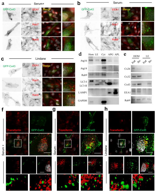
Figure 5. Internalized Cx43 is targeted to recycling endosomes during serum deprivation.
(a–c) Immunofluorescence for the indicated endosomal markers in NRK cells expressing GFP-Cx43 maintained in presence of serum (a), absence of serum (b) or treated with lindane (c). Single black and white inverted channels, merged channels and higher magnification insets are shown. (d) Immunoblots for indicated proteins in isolated fractions from starved mouse liver HOM: homogenate; LE: late endosomes; CYT: cytosol; APG: autophagosomes; APL: autophagolysosomes. (e) Immunoblots for different connexins in homogenates and late endosomes isolated from fed and 6h starved mice. (f–h) Fluorescence images of NRK cells expressing GFP-Cx43 maintained in presence of serum (f), absence of serum (g) or treated with lindane (h) and incubated with Alexa594-transferrin for 15 minutes before fixation. Top: single and merged channels and boxed area at higher magnification. Bottom: 3D reconstructions and higher magnification details of the boxed areas. Nuclei are highlighted with DAPI. Bars: 5 μm. Uncropped images of blots are shown in Supplementary Fig. 9.

