
Fig. 1.
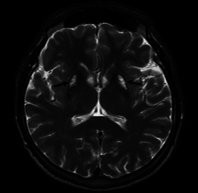
The MR image mimicking the “eye of the tiger” sign in T2-weighted magnetic resonance sequences; bilateral hypointensity in both putaminal regions and lateral parts of globi pallidi, with a central hyperintensity (black arrows)
Official websites use .gov
A
.gov website belongs to an official
government organization in the United States.
Secure .gov websites use HTTPS
A lock (
) or https:// means you've safely
connected to the .gov website. Share sensitive
information only on official, secure websites.
The MR image mimicking the “eye of the tiger” sign in T2-weighted magnetic resonance sequences; bilateral hypointensity in both putaminal regions and lateral parts of globi pallidi, with a central hyperintensity (black arrows)