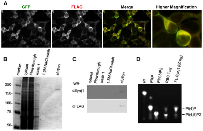
Fig. 1. Expression and purification of active Synj1 from HEK-239T cells.

(A) HEK-293T cells stably expressing Synj1-GFP-FLAG labeled with indicated antibodies. Image was acquired with a 40x objective on an Olympus IX81 microscope using image analysis software Slidebook version 5.0. (B) Commassie stained gel showing aliquots from purification scheme of Synj1-GFP-FLAG. (C) Western blot detection of Synj1-GFP-FLAG using Synaptojanin 1 and FLAG specific antibodies. (D) Purified Synj1-GFP-FLAG phosphatase activity was detected using BODIPY labeled lipids and a positive control for lipid phosphatase activity, rat brain cytosol (RBC).
