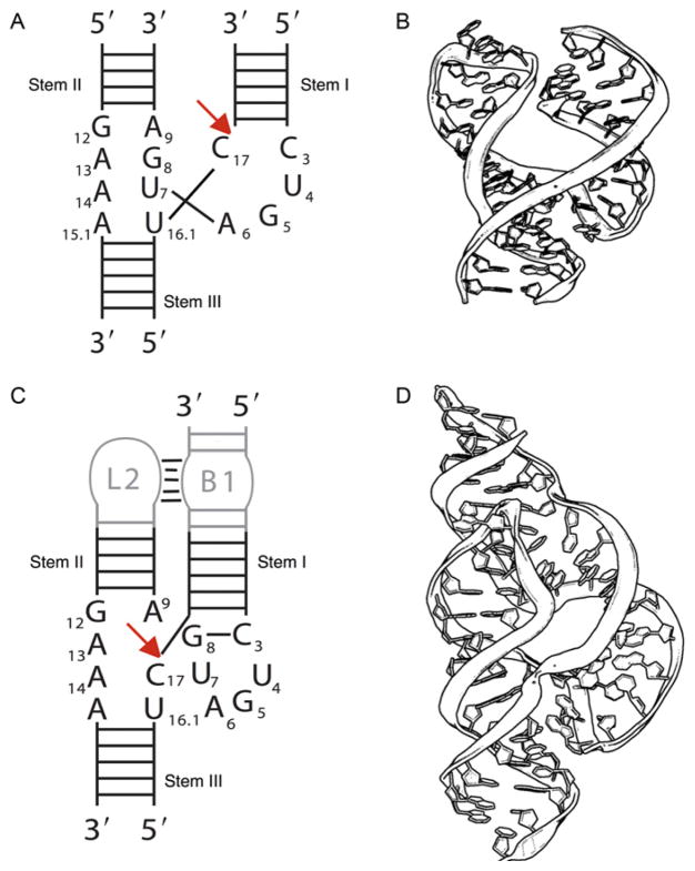
Figure 1.1.
The minimal and full-length hammerhead ribozymes. (A) A schematic representation of the secondary structure of the minimal hammerhead ribozyme. (B) The crystal structure of a corresponding minimal hammerhead ribozyme. The longer strand is the enzyme and the shorter strand is the substrate. (C) A schematic representation of the full-length hammerhead ribozyme emphasizing the presence of a tertiary contact between stem(s) I and II. (D) The crystal structure of a corresponding full-length hammerhead ribozyme. Again, the longer strand is the enzyme and the shorter strand is the substrate.

