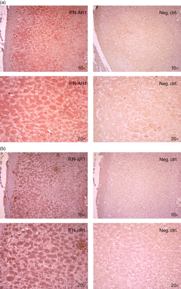
Figure 2.

Expression of interferon alpha/beta receptor chain 1 (IFN-AR1) and IFN lambda receptor 1 (IFN-λR1) in human adrenal cortex. Human adrenal tissue sections were stained with (a) anti-human IFN-AR1 or (b) anti-human IFN-λR1 antibodies followed by horseradish peroxidase (HRP)-conjugated secondary antibodies. Positive structures were visualized with 3-amino-9-ethylcarbazole substrate (red) and cell nuclei counterstained with haematoxylin (blue). Neg. ctrl = staining performed with secondary antibody only. Images were obtained with ×10 and ×20 objectives as indicated.
