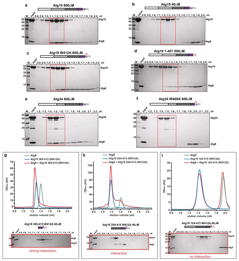
Figure 1.
Atg19 and Atg34 interaction with Atg8
(a-f) Atg8 was co-incubated with Atg19 or Atg34 (wild type and mutants) at concentrations of 40μM and 800μM, respectively and run on a size exclusion column. Aliquots of individual fractions were run on SDS-PAGE gels and stained by Coomassie. The white box indicates the N-terminal domain, the light grey box the coiled-coil domain, the dark grey box the Ams1-binding domain and the purple box indicates the C-terminal domain. (g-f) Atg8 was co-incubated with the indicated Atg19 domains at 40 μM and run on a size exclusion column. Individual elution profiles are shown above; Coomassie stained SDS-PAGE gels below. Columns: (a-f, i: S200 3.2/30; g, h: S75 3.2/30). M: size marker. All experiments shown have been done two times.

