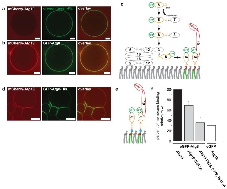
Figure 4.
Reconstitution of Atg8-dependent Atg19 recruitment to the membrane
(a) GUVs incubated with mCherry-Atg19. The membrane was marked by incorporation of an Oregon green labelled lipid. (b) GUV coated with GFP-Atg8 incubated with mCherry-Atg19. (c) Scheme showing the proteins and reaction resulting in GFP-Atg8 conjugation to the GUV shown in (b). (d) GUVs containing nickel lipids incubated with GFP-Atg8-6xHis and wild type mCherry-Atg19. (e) Experimental setup of the experiment shown in (d). (f) Quantification of membrane binding by the indicated mCherry-Atg19 proteins. The experimental setup is shown in (e). The graph is based on 3 experiments. Shown are the averages and s.d. of these 3 experiments (N = 3, based on 318 GUVs for Atg19, 171 GUVs for Atg19 W412A, 166 GUVs for Atg19 F376A, F379A, W412A, 188 GUVs for the eGFP control). Scale bars: 5μm. Abbreviations: 3: Atg3; 5: Atg5; 7: Atg7; 8: Atg8; 12: Atg12; 16: Atg16; 19: Atg19. Experiments shown in (a, b) have been conducted two times; the experiment in (d) three times.

