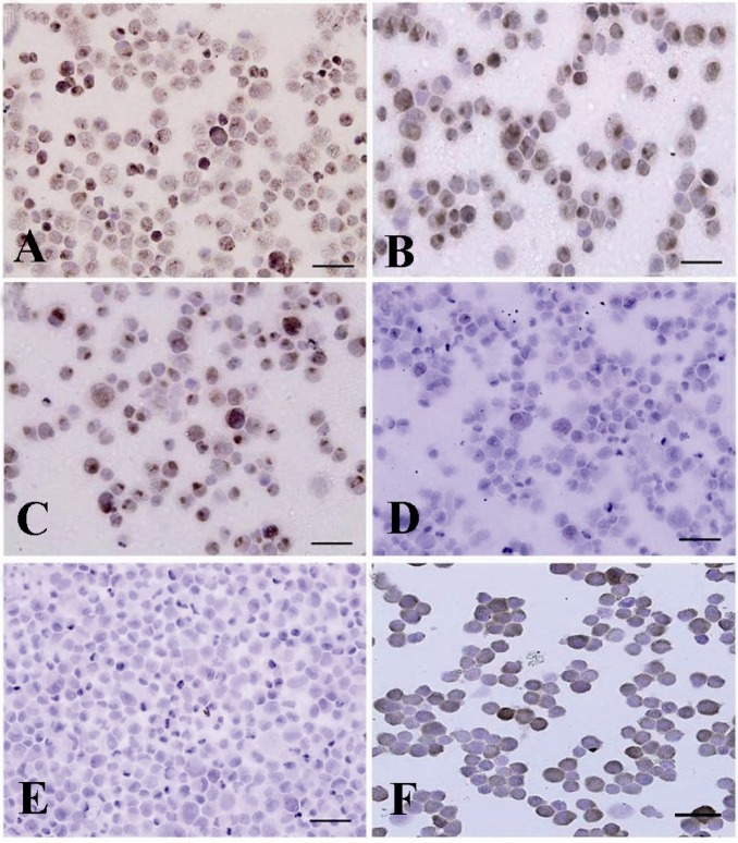
Figure 3.
Immunocytochemical identification of undifferentiated type A spermatogonia after percoll separation method using an antibody against PGP9.5. Basal spermatogonia (dark brown) and some aggregated spermatogonia (light brown) in gradients A) 32% and B) 30% were significantly higher than C) 28% and D) 20% gradients. Note the intense staining in the cytoplasm, especially in the vicinity of the nucleus, in basal spermatogonia. E) Negative and F) positive controls were detected

