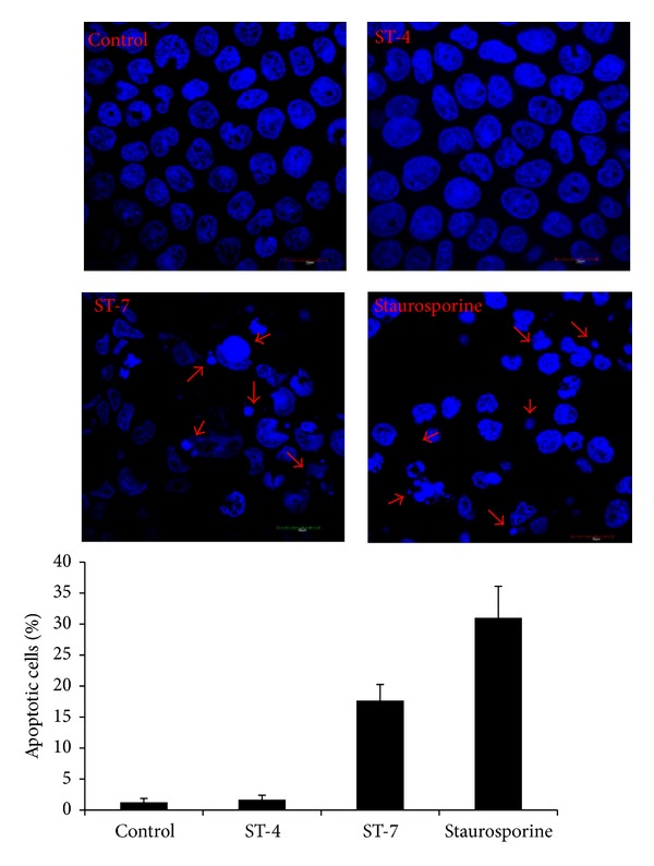
Figure 6.

Representative fluorescence micrographs showing apoptosis of Caco-2 cells after DAPI staining. Cells were grown on glass coverslips and incubated for 24 h with culture media, ST-4, ST-7, or 0.5 μM staurosporine as positive control. Cells coincubated with ST-7 and staurosporine exhibit nuclear fragmentation and condensation (arrow) typical of apoptotic cells. Histogram represents percentage of apoptotic cells after DAPI fluorescence assay. Caco-2 monolayers coincubated with ST-7 exhibited significantly higher percentage of apoptosis (P value < 0.05) compared to ST-4 and negative control. Values are means ± standard error (n = 6 per group). For each sample, ~100 cells were counted at 1000x magnification.
