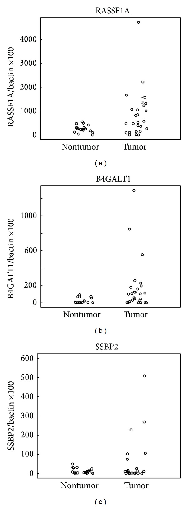
Figure 3.

Quantitative MSP results of hepatocellular carcinoma samples and adjacent normal liver samples that were bisulfite treated to examine the promoter methylation status of RASSF1A, B4GALT1, and SSBP2. Scatter plots of quantitative MSP analysis of candidate gene promoters. Twenty-two adjacent normal liver tissue samples and 27 hepatocellular carcinoma samples were tested for methylation for each of the three genes by quantitative MSP. The relative level of methylated DNA for each gene in each sample was determined as a ratio of MSP for the amplified gene to ACTB and then multiplied by 100 for easier tabulation (average value of duplicates of gene of interest/average value of duplicates of ACTB × 100). The samples were categorized as unmethylated or methylated based on detection of methylation above a threshold set for each gene. This threshold was determined by analyzing the levels and distribution of methylation, if any, in normal, age-matched tissues.
