Figure 6.

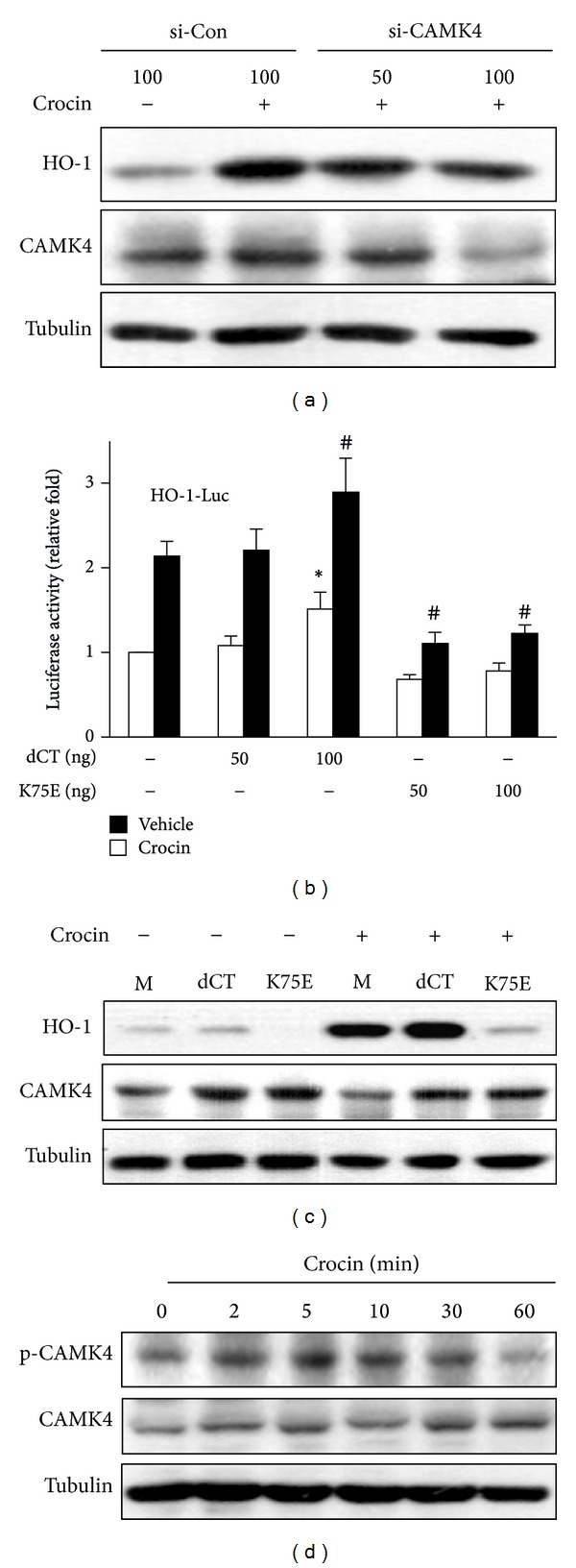
Involvement of CAMK4 in crocin-mediated expression of HO-1. (a) RAW 264.7 cells were transfected with CAMK4 siRNA or control siRNA and then treated with crocin (500 μM) for 6 h. Equal amounts of cytosolic proteins were analyzed by Western blotting. (b) Cells were cotransfected with HO-1 promoter-luciferase construct and pcDNA3.1, CAMK4 constitutive active (dCT), or kinase dead (K75E) construct and then treated with crocin (500 μM). Equal amounts of cell extracts were assayed for dual luciferase activity. *P < 0.05 versus the pcDNA3.1-transfected (vehicle-treated) group; # P < 0.05 versus the pcDNA3.1-transfected (crocin-treated) group. (c) Cells were transfected with pcDNA3.1 (mock, M), dCT or K75E construct and then treated with crocin (500 μM). Equal amounts of cytosolic proteins were analyzed by Western blotting. (d) Cells were treated with crocin (500 μM) for the indicated time and the levels of phosphorylated CAMK4 and total CAMK4 were analyzed by Western blotting. Tubulin was used as a loading control.
