
Figure 6.
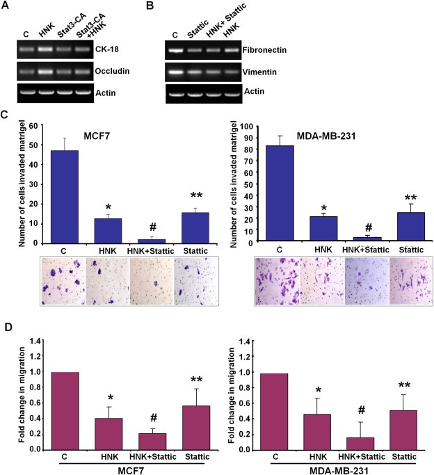
Stat3‐inhibition plays an important role in honokiol‐mediated modulation of EMT markers, and inhibition of invasion and migration of breast cancer cells. A, MCF7 cells were treated with vehicle (C) or 5 μM honokiol (HNK), transfected with constitutively active Stat3 (Stat3‐CA) and treated with Honokiol (Stat3‐CA + HNK). Total RNA was isolated and subjected to RT‐PCR analysis using CK‐18 and occludin primers. Actin was included as control. B, MDA‐MB‐231 cells were treated with vehicle (C), 10 μM Stattic, 5 μM honokiol (HNK) alone, or in combination (HNK + Stattic), total RNA was isolated and subjected to RT‐PCR analysis using fibronectin and vimentin primers. Actin was included as control. C, MCF7 and MDA‐MB‐231 cells were cultured in matrigel‐invasion chambers followed by treatment with 10 μM Stattic, 5 μM honokiol (HNK) alone, or in combination (HNK + Stattic) for 24 h as indicated. C represents vehicle controls. The number of cells that invaded through the matrigel was counted in five different regions. *P < 0.005, compared with vehicle‐treated controls; **P < 0.001, compared with vehicle‐treated controls; #P < 0.005, compared with HNK‐treated cells. D, MCF7 and MDA‐MB‐231 cells were subjected to spheroid‐migration assay. Culture media were replaced with media containing 10 μM Stattic, 5 μM honokiol (HNK) alone, in combination (HNK + Stattic) or vehicle control (C). The spheroids were photographed 48 h‐post treatment. The results are shown as fold‐change in migration of breast cancer cells in response to HNK and Stattic treatments. These are representative of three independent experiments performed in triplicates. *P < 0.01, compared with vehicle‐treated controls; **P < 0.05, compared with vehicle‐treated controls; #P < 0.05, compared with HNK‐treated cells.