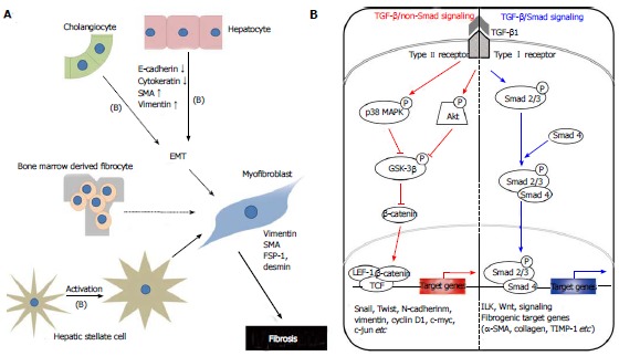
Figure 1.

Mechanisms of hepatic fibrogenesis. A: The proposed sources of hepatic myofibroblasts: Resident cells (hepatic stellate cells and portal fibroblasts); bone marrow-derived mesenchymal cells, and EMT from hepatocytes and cholangiocytes. Different insults initiate inflammation and then cause hepatocyte stellate cells activation and hepatocyte and biliary cell damage, necrosis and EMT. Continuous insults will shift those EMT-like cells to complete EMT cells and finally myofibroblasts, the main producer of extracellular matrix, which may be one of the main causes of an early loss of regenerative capacity. A similar process also occurs in biliary cells. Some cytokines play an important role to affect the adjacent cells and promote EMTs, such as TGF-β1 (B); B: Schematic presentation of the major intracellular signal transduction pathways of TGF-β1 in liver fibrosis. TGF-β1 is a chief inducer of the EMT process, and p-Smad2/3, p38 MAPK and ILK function as mediators of the intracellular signaling pathway. TGF-β1 signals via heteromeric transmembrane complexes of type I and type II receptors that are endowed with intrinsic serine/threonine kinase activity (ALK activin receptor-like kinase). Upon type-II-mediated phosphorylation of the type I receptor, the activated type I receptor initiates intracellular signalling by phosphorylating receptor regulated-Smad2 and Smad3. Activated Smads form heteromeric complexes with Smad4 and these complexes accumulate in the nucleus where they mediate transcriptional responses. p-Smad2/3: Phosphorylated-Smad2/3; MAPK: Mitogen-activated protein kinase; GSK-3β: Glycogen synthase kinase-3β; ILK: Integrin-linked kinase; TCF/LEF-1 complex: T cell factor/lymphoid enhancer-binding factor-1 complex; EMT: Epithelial-mesenchymal transition; TGF: Transforming growth factor.
