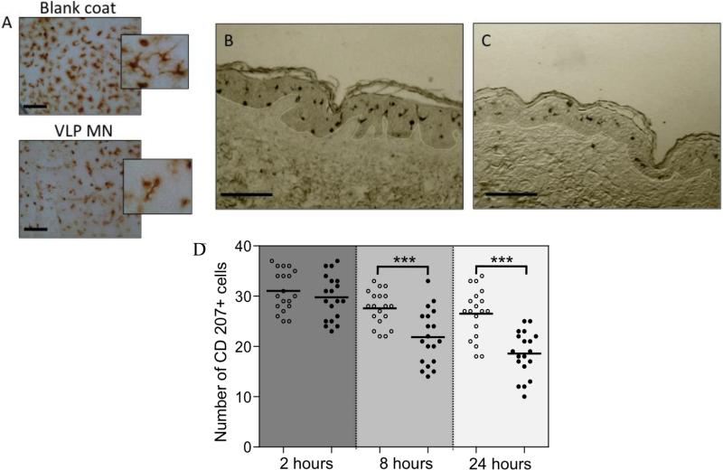
Figure 5. Morphological and migratory changes to dendritic cells following vaccine delivery.
Immunohistochemical study of LCs in human skin following microneedle delivery of H1N1 VLPs. A) Epidermal sheets isolated from skin 24 h after placebo (Blank coat) and VLP (VLP MN) delivery (Bar = 100μm). B, C) Immunohistochemically stained longitudinal sections of human skin treated with (B) VLP and (C) placebo (Bar = 200μm). D) Immuno-stained epidermal sheets were visualised by light microscopy 2, 8 and 24 h after microneedle delivery of H1N1 VLPs. The number of CD207+ cells observed in 200μm2 fields of view are shown for VLP treated samples (black circles) and placebo treated samples (white circles). A total of 3-5 replicate counts were taken from a total of 4 independent skin donors. ***denotes significant (P<0.01) difference between VLP and control samples at each timepoint.

