
Figure 1.
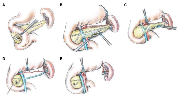
Tumors of the pancreatic head. A: A pancreatic neuroendocrine tumor (PNET) involving the posterior aspect of the pancreatic head, after adequate exposure by extensive kocherization and medial retraction of the pancreatic head, prior to enucleation; B: A PNET involving the inferior border of the body/tail of the pancreas. Resection is being performed using the LigaSure device; C: PNETs located in the posterior aspect of the body/tail occasionally require partial resection of the splenic vein in order to perform successful enucleation; D: The intraoperative appearance after performance of spleen-preserving distal pancreatectomy with splenic vessel preservation; E: The intraoperative appearance after performance of spleen-preserving distal pancreatectomy without splenic vessel preservation. Figure 1D and E represent patients with multiple endocrine neoplasia-1, with an additional PNET located in the head. This synchronous tumor will be excised by enucleation.