
Figure 1.
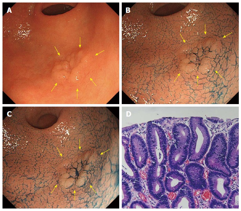
A case of tubular adenoma (no change in acetic acid indigocarmine mixture-chromoendoscopy). A: Whitish superficial elevated lesion is shown at the greater curvature of the antrum in white light endoscopy (indicated by yellow arrows); B: After sprinkling indigo carmine solution; C: 3 min after sprinkling acetic acid indigocarmine mixture (AIM) solution. Compared to B, there was no surface color change (no change in AIM-chromoendoscopy); D: Histology after endoscopic submucosal dissection.