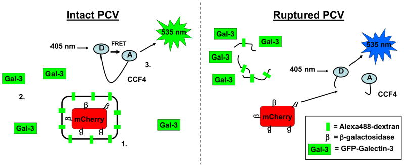
Fig 2. Detection of vacuole rupture.
Three recent techniques allow the detection of vacuole rupture in real time on a single cell basis. (1) Alexa488-Dextran marks the vacuole membrane and colocalization with bacteria expressing mCherry can be detected by fluorescence microscopy. (2) GFP-tagged Galectin-3 is specifically recruited to the remnants of ruptured vacuoles. (3) Following vacuole disruption, surface-expressed β-lactamases can cleave cytosolic CCF4. Cleavage of CCF4 relieves the intramolecular FRET and causes a change in fluorescence emission.

